 |
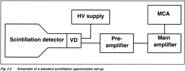
| Pulse Height Spectroscopy The basic principle of pulse height spectroscopy is that the light output of a scintillator is proportional to the energy deposited in a crystal. The standard way to detect scintillation light is to couple a scintillator to a photomuitiplier. Furthermore, a g-ray spectrometer usually consists of a preamplifier, a main (spectroscopy) amplifier and a multichannel analyzer (MCA). The electronics amplify the PMT charge pulse resulting in a voltage pulse suited to detect and analyze with the MCA. The schematic is shown in Fig. 2.2. For a typical pulse height spectrum see Fig. 2.1. |
|||||||||||
 |
|||||||||||
| BACK TO TOP | |||||||||||
| Energy Resolution An important aspect of a g -ray spectrometer is the ability to discriminate between g-rays with slightly different energy. This quality is characterized by the so-called energy resolution which is defined as the width (FWHM) of the photopeak at a certain energy. Besides by the g -ray energy, the energy resolution is influenced by :
At low energies where photoelectron statistics dominate the energy resolution, the energy resolution is roughly inverse proportional to the square root of the g-ray energy. The energy resolution of a scintillation detector is a true detector property, limited by the physical characteristics of the scintillator and the PMT or other readout device. A typical energy resolution for 662 keV g-rays absorbed in small NaI(Tl) detectors is 7.5 % FWHM. At low energies, e.g. at 5.9 keV, a typical value is 45 % FWHM. At these low energies, surface treatment of the scintillation crystal strongly influences the resolution. It may be clear that especially at low energies, scintillation detectors are low resolution devices unlike Si(Li) or HPGe detectors. |
|||||||||||
| BACK TO TOP | |||||||||||
|
The time resolution of a scintillation detector reflects the ability to define precisely the moment of absorption of a radiation quantum in the detector. The light pulse of a scintillator is characterized by a rise time and by a 1/e fall time t (= decay time see section 3.1). It is obvious that the best time definition of an absorption event is obtained when the scintillation pulse is short (small decay time) and intense. Furthermore, the rise time and time jitter of the PMT and of the electronics are important. Small cm size NaI(Tl) detectors have typical time resolutions of a few nanoseconds for 60Co (1.2 MeV). Much better time resolutions can be obtained with plastic or BaF2 scintillation crystals. BaF2 is presently the fastest known scintillator with detector time resolutions of a few hundred picoseconds. |
|||||||||||
| BACK TO TOP | |||||||||||
| Peak-to-valley
Ratio A sensitive way to check the energy resolution of a scintillation detector is to define a so-called peak-to-valley (P/V) in the energy spectrum. This criterium does not depend on any possible offsets in the signal. Either the peak-to-valley between two gamma peaks is taken or the ratio between a low energy peak and the PMT / electronic's noise. A good P/V ratio for a 76 x 76 mm NaI(Tl) crystal is 10 : 1. This is equivalent to an energy resolution of 7.0 % at 662 keV. At 5.9 keV, a high quality X-ray detector can have a P/V ratio of 40 : 1. |
|||||||||||
| BACK TO TOP | |||||||||||
| Spectrum Stabilization Extreme count rate changes, temperature variations or instable electronics may cause peak position variations in a spectrum. To compensate for these effects it is possible to calibrate the peak position with a so-called Am-pulser . This is a very small radioactive 241Am source mounted inside a scintillation detector. The a-particles, emitted by the 241 Am, cause scintillations in the crystal that are detected by the PMT (or the photodiode) of the detector. For NaI(Tl), the a -peak is situated between a Gamma Equivalent Energy (GEE) of 1.5 and 3.5 MeV and can be specified. Count rates are 50, 200 and 1000 cps. The position of the pulser peak is used as a reference to compensate for the above mentioned variations in detector response. The above way of calibration is not ideal since the response of most scintillation crystals for g-rays and a-particles is different. However, a second order compensation using e.g. a thermistor is only necessary for large temperature ranges. Also other spot-activated scintillation crystals can be used for the above application. The best choice depends on the type of host crystal, the required GEE and required calibration accuracy. For occasionally monitoring your system integrity, Light Emitting Diodes (LEDs) or laser ports can also be used. LEDs can be mounted inside scintillation detectors or a window for that purpose can be provided. Apart from these ways of pulse height stabilization, it is of course possible to stabilize electronically on the peak of an (always present) external source. Sometimes the 40K background line can be used for this purpose. |
|||||||||||
| BACK TO TOP | |||||||||||
| General Information Interactions in Scintillation Materials Scintillation Response to g-rays Scintillator Interaction with Charged Particles; a - and b-particle detection |
|||||||||||
| BACK to Scintillation Detectors | |||||||||||